Lets first talk about the alignment of the native knee, then compare it with the goals for TKA alignment.
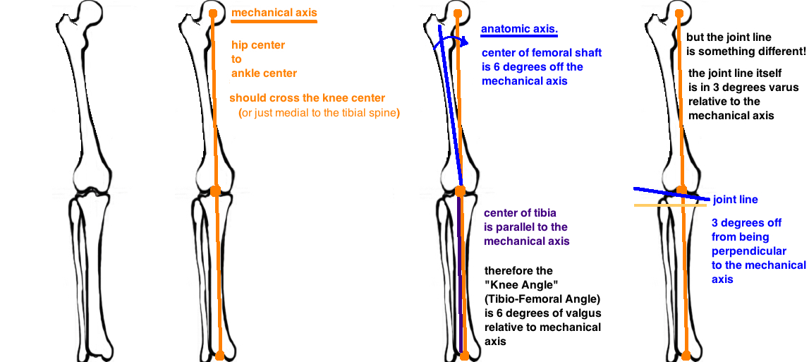
The Mechanical Axis of the leg is a line drawn from the center of the hip to the center of the ankle. This line should cross through the center of the knee. This means that the knee has neutral alignment.
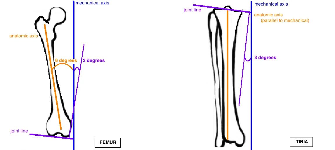
The Anatomic Axis is the center of the bones making up the leg, and the anatomic axis of the femur is 6° from the mechanical axis while the anatomic axis of the tibia is inline with the mechanical axis. Therefore the Knee Angle (referring to the Femoral-Tibial Angle: FTA) is 6° valgus (relative to the mechanical axis).
We also need to consider the joint line itself. The joint line is variable person-to-person, however, on average its in about 3° of varus (relative to the mechanical axis). On the tibial side, it means the tibial articular surface is in 3° varus, while on the femoral side, the femoral articular surface is in 3° of valgus. Now we need to combine everything together. On the femoral side, the joint line is in 9° of valgus relative to the anatomic axis (thats 6° from the femoral center to the mechanical axis and then 3° more from the mechanical axis to the line across the distal femoral condyle). On the tibial side, the joint line is in 3° of varus relative to the anatomic axis (thats 0° from the tibial center to the mechanical axis because these two are parallel, and then 3° from the mechanical axis to the tibial joint line).
In a standard TKA (we describe in more detail below) these angles are simplified by cutting the tibia perpendicular to the mechanical/anatomic axis, which is 0°. All of the femoral cuts are then adjusted to line up with this tibial cut.
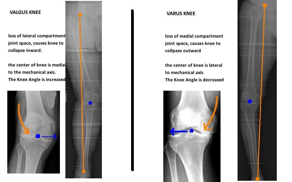
Knee deformity.
A Varus knee (due to common medial sided DJD) will deviate laterally with respect to the mechanical axis due to narrowing of the medial compartment and gradual attenuation of the lateral soft tissue.
A Valgus knee will deviate medially (the knock-kneed appearance on clinical exam) due to narrowing of the lateral compartment and attenuation of the medical soft tissue. If the compartments are equal, and there is no gross bony deformity, the knee should not be angulated and should remain centered within the mechanical axis.